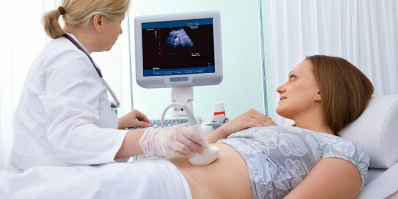
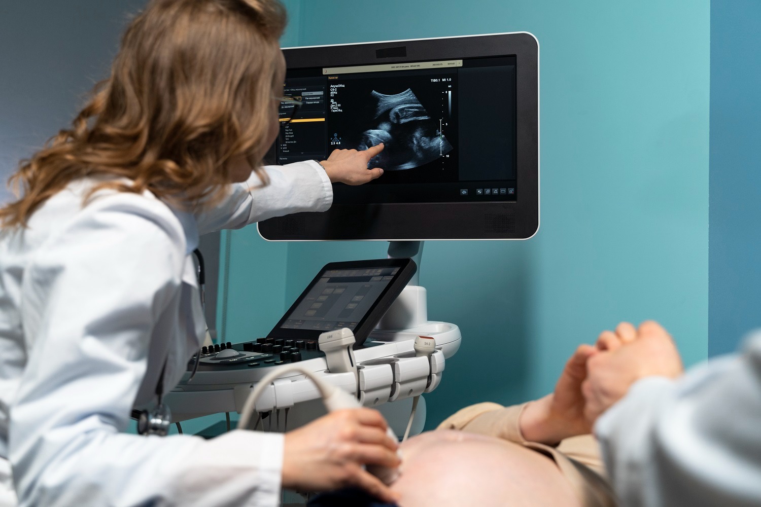
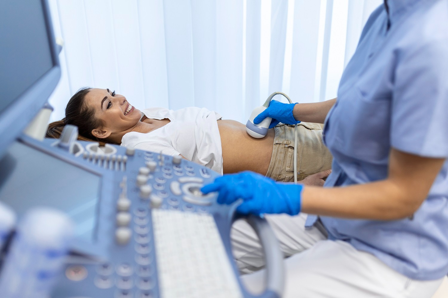
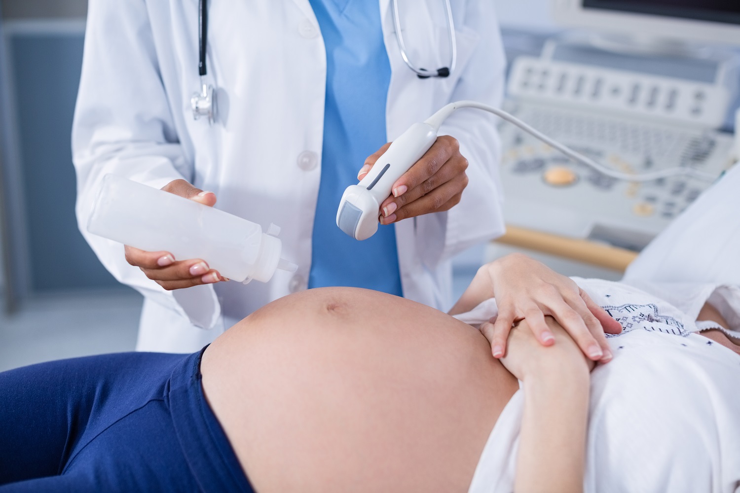
High-frequency sound waves are referred to as ultrasounds. These sound waves are used in ultrasound exams to create an image or picture of your inside organs on a screen. A radiologist performs an ultrasound by moving a smooth, handheld instrument known as a transducer in a sliding and rotating motion across the body. The high-frequency sound waves enter your body through the transducer. Different soft tissues, structures, or bodily parts reflect different sound waves in different ways. A moving image is produced on a screen by converting these sound waves into electrical impulses.
There are several benefits to an ultrasound. It is extremely safe because it causes no discomfort and doesn’t use radiation. The high-frequency sound waves guarantee that photographs display a great deal of detail, allowing one to see even the smallest bodily components. Ultrasound is great for imaging infants and youngsters since it may be performed while the subject is moving.
An ultrasound may be requested for a variety of reasons. It is used, for instance, to inspect the abdominal organs and other body components. You may use colour Doppler ultrasonography to observe blood flow in any vein or artery in any area of your body. The musculoskeletal system, which includes the muscles, bones, and joints, may be assessed using high-resolution ultrasound. An essential component of evaluating any breast mass is breast ultrasonography. Ultrasound is a great diagnostic technique since it can capture high-quality pictures of the majority of your body.
The kind of ultrasound that is sought will determine this. Some of the common tests are listed below, and preparation is usually necessary.
Dress in a way that will allow you to easily enter the area being photographed.
For comparison and evaluation, bring any prior radiological exams you have had, such as computed tomography scans, X-rays, magnetic resonance imaging, and ultrasound, with you.
Abdomen ultrasound: 4-6 hours before to the test, you will often need to fast (not eat or drink anything). This guarantees that the region to be checked is free of food or liquid. Additionally, it makes sure the gall bladder is enlarged for a better view.
Breast ultrasonography: No prior preparation is necessary.
This ultrasound scan looks at the female pelvic organs. This comprises the uterus or womb, the ovaries, the cervix, and the endometrium, which is the lining of the uterus.
It is advised to use both transvaginal and transabdominal scans to obtain sufficient images of your pelvic tissues. The process of transabdominal scanning involves applying a probe and gel to your lower abdomen’s skin. This offers a decent general evaluation of the pelvic organs without going into great depth. During a transvaginal scan, a sterile, covered probe is carefully inserted into the vagina while you are still covered. Because this probe is positioned closer to the uterus, ovaries, and cervix, it often produces crisper pictures of the pelvic tissues. A PAP smear is more unpleasant than this. You will always be in control of whether or not this is done.
Clearer pictures can be acquired during transabdominal scanning when there is fluid in the bladder. It facilitates better visualisation by removing your intestine from the pelvis and creating a “window” for the scan. Partially filling your bladder is necessary; it shouldn’t be so full that it hurts. It is recommended that you drink half a litre of water prior to your appointment and empty your bladder two hours prior to the examination. Before the transvaginal scan starts, you will be given the opportunity to empty your bladder.
There is no need for preparation for thyroid ultrasonography.
Ultrasound test: No prior preparation is necessary.
There is no need to prepare for musculoskeletal (relatingto muscles, bones, and joints) ultrasonography.
An hour prior to the renal (kidney-related) ultrasonography, you must consume 750 millilitres of water. After consuming the liquids, avoid using the loo. The bladder will swell after drinking the water, making it possible to view it and the surrounding internal organs.
Renal (kidney) artery ultrasound: To make sure that the renal arteries are not obstructed by food or liquid, you must fast for eight hours before to the test.
Ultrasound is a safe examination that provides excellent imaging without any significant risk to the patient.
In addition to being a safe process without the hazards associated with radiation-based imaging, ultrasound offers superb imaging of the body’s soft tissues. When performed in a suitable clinical context, such as a hospital or private radiology practice, there are no known negative consequences of sound waves at the levels employed in ultrasonography.
Ultrasound is perfect for imaging infants and youngsters since it may be utilised while the patient is moving. In musculoskeletal (related to muscles, bones, and joints), gynaecological (related to women’s health, particularly the reproductive organs), and vascular (related to blood vessels) ultrasonography, imaging movement is also highly helpful. The ability to view the inside of the body in postures or with motions where there is discomfort or mobility restriction is made possible by dynamic imaging, or moving pictures, which are produced by images employing ultrasonic sound waves.
In rare cases, a particular ultrasound contrast agent is injected into an arm vein to identify certain illnesses or issues. The radiologist will explain this to you during the examination if they think it would be helpful.