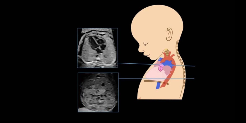
The evaluation of the baby’s heart before to delivery is known as foetal echocardiography. The majority of kids have healthy hearts. As a result, the majority of prenatal heart scans reveal no clear anomalies, and the main cardiac structures are discernible as they would be in a healthy foetus.
About 8 out of every 1,000 live births will have a cardiac abnormality. If a specialist scan of the foetal heart is conducted, half of these problems can be detected before the baby is born. The majority of complicated defects are easily tolerated by the infant throughout pregnancy, but because of changes in the way the blood circulates after delivery, therapy is typically necessary after birth and possibly as early as the first few days of life.
Even after birth, minor anomalies such tiny holes in the heart may be found, but they do not impair the heart’s function. Minor abnormalities often don’t require treatment, but when necessary, follow-up may be required. Certain newborns will also need to be evaluated for rhythm abnormalities in addition to structural issues.
Foetal echocardiography is often performed between weeks 22 and 24 of pregnancy, and one scan is usually sufficient to evaluate the foetal heart. But in certain cases, the foetal heart may be evaluated as early as 13–14 weeks of pregnancy, which gives some parents who are worried about having a child with a congenital heart problem a lot of knowledge and comfort. Later in pregnancy, a second foetal echo is planned in these situations.
Before the test, you are not need to drink anything or have your bladder full.