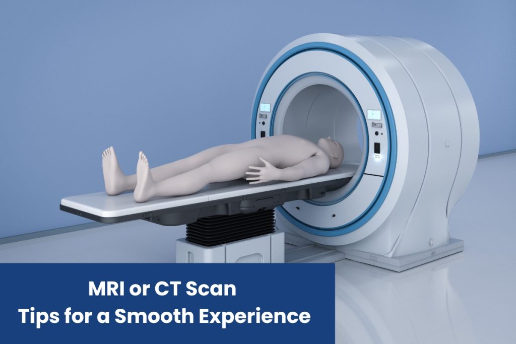
Preparing for an MRI or CT Scan: Tips for a Smooth Experience
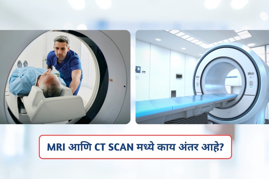
Medical imaging procedures like MRI (Magnetic Resonance Imaging) and CT (Computed Tomography) scans are essential diagnostic tools that help doctors identify and treat various health conditions. While these scans are non-invasive and painless, preparing for them can feel overwhelming, especially if it’s your first time. Proper preparation ensures a smooth experience and accurate results. Here are some practical tips to help you get ready for your MRI or CT scan. 1. Understand the Procedure Before your scan, take time to understand what the procedure entails. MRI Scan: Uses powerful magnets and radio waves to create detailed images of your body. It’s commonly used for examining soft tissues, the brain, joints, and organs. CT Scan: Uses X-rays to produce cross-sectional images of your body. It’s often used for detecting bone fractures, tumors, and internal injuries. Knowing what to expect can ease anxiety and help you feel more in control. 2. Follow Pre-Scan Instructions Your healthcare provider or diagnostic center will provide specific instructions. These may include: Fasting: For some scans, you may need to avoid eating or drinking for a few hours beforehand. Medications: Inform your doctor about any medications you’re taking. You may need to adjust or pause them before the scan. Clothing: Wear comfortable, loose-fitting clothing without metal zippers, buttons, or jewelry. You may be asked to change into a hospital gown. Contrast Dye: If your scan requires a contrast dye, let the technician know about any allergies or kidney problems. 3. Communicate with Your Technician Don’t hesitate to share any concerns or questions with the technician before the scan. Inform them if you: Have claustrophobia (fear of enclosed spaces). Are pregnant or suspect you might be. Have any implants, such as pacemakers or metal prosthetics. Experience pain or discomfort in certain positions. 4. Stay Calm and Relaxed It’s natural to feel nervous, but staying calm can make the process easier. Practice Deep Breathing: Slow, deep breaths can help reduce anxiety. Listen to Music: Some centers allow you to listen to calming music during the scan. Close Your Eyes: If you’re uncomfortable in the MRI machine, closing your eyes can help. 5. Plan for Post-Scan Recovery Most MRI and CT scans don’t require recovery time, but if you’ve had sedation or contrast dye, you may need to rest for a few hours. Arrange for someone to drive you home if necessary. Why Choose Inamdar MRI and CT Scan Diagnostic Center? Advanced Technology: Equipped with cutting-edge MRI and CT scan machines for precise diagnostics. Expert Team: Skilled radiologists and technicians who prioritize patient care. Comfortable Environment: A welcoming atmosphere designed to put patients at ease. Quick Results: Timely and accurate reports to aid in your treatment plan. Preparing for an MRI or CT scan doesn’t have to be stressful. By following these tips and choosing a reliable diagnostic center like Inamdar MRI and CT Scan Diagnostic Center, you can ensure a smooth and hassle-free experience. Remember, these scans are a vital step in maintaining your health, so take the time to prepare properly and trust the experts to guide you through the process. If you’re in need of an MRI or CT scan, contact Inamdar MRI and CT Scan Diagnostic Center today to schedule your appointment and experience top-notch diagnostic care.
Preparing for an MRI or CT Scan: Tips for a Smooth Experience Read More »